Breast Imaging скачать fb2
Beverly Hashimoto - Breast Imaging краткое содержание
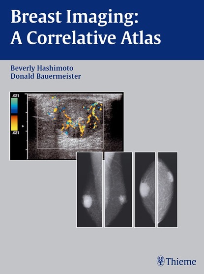
An extensive armamentarium of imaging techniques can help you accurately detect all breast lesions and avoid missed diagnoses. This book greatly expands your options by integrating both mammographic and sonographic findings, with more than 200 case examples covering a wide range of clinical conditions. Its unique format–organized by two distinct tables of contents–helps you locate a specific topic by either mammographic lesion or sonographic image.The book highlights the importance of high-resolution sonography and its cross-correlation with mammography by providing a broad spectrum of imaging entities, from common pathologies to rare breast abnormalities. You'll also get helpful information on normal anatomy, new technology, instrumentation, and management issues.Special features of this state-of-the-art resource:Provides clear visual patterns–with more than 700 detailed illustrations–that help you identify both mammographic and sonographic abnormalitiesPresents more than 200 cases showing a variety of scenarios, all cross-referenced for easy useIncludes two different tables of contents organized by both mammographic and sonographic findings–for easy and quick referenceGives you pearls and pitfalls describing your clinical and radiographic options for even the most unusual casesOffers magnified views of calcification lesions for clear demonstration of conceptsHere is the quintessential imaging atlas that radiologists, residents, and other physicians need to accurately integrate mammographic and sonographic findings. Benefit from these unique diagnostic tools that will upgrade and refine your skills in virtually any clinical situation involving breast imaging!
Скачать книгу «Breast Imaging» Beverly Hashimoto
Чтобы оставить свою оценку и/или комментарий, Вам нужно войти под своей учетной записью или зарегистрироваться


